Amyotrophic lateral sclerosis (ALS) is characterised by progressive degeneration of upper (UMN) and lower (LMN) motor neurons in the brain and spinal cord. Rare in its own right, ALS is the most common form of motor neuron disease (MND). Primary lateral sclerosis, a disease isolated to UMNs, makes up 1–3 %, and progressive muscular atrophy, limited to LMNs, approximately 10 % of MND. Predominance of UMN features probably carries a better prognosis, even for patients with ALS.1 Incidence rates for ALS range from 1.2–4.0 per 100,000 person years in Caucasians.2–5 The rate may be lower in some minority populations6 and historically as much as 100 times higher in the western Pacific (Guam, Japan’s Kii Peninsula, western New Guinea).7Incidence increases with age, peaking between 70 and 80 years and is thought to be more common in men than women.8,9
ALS begins in limb or bulbar muscles and then spreads to contiguous andeventually respiratory, myotomes. Survival ranges from months to decades, but is usually less than 3 years from when symptoms first appear.10
Jean-Martin Charcot, the father of neurology, used the clinicoanatomical method that he devised to describe the clinical and pathological features of ALS during series of lectures in the 1860s and 1870s.11 The methodology available to Charcot was primitive by today’s standards, but his approach was so insightful, becoming the foundation of the field he formed, that his original descriptions are considered accurate today. Sir William Gowers and Lord Russell Brain made major contributions in the UK, labelling the malady MND instead of ALS because they believed that all patients have pathology of both UMNs and LMNs, either during life or at autopsy. Few American neurologists early in the 20th Century meant that the illness was largely overlooked in the New World until 1939 when the notable first baseman for the New York Yankees was diagnosed with ALS. Called the “iron horse” for playing 2,130 consecutive games over 14 years, Gehrig first exhibited signs in 1938.12,13 Several weeks into the 1939 season, he removed himself from the lineup, no longer able to play. Gehrig died two years later, having participating in an early clinical trial.14 ALS still uses Gehrig’s name as its eponym in the US; MND is used in the UK, whereas ALS is preferred in France and the US.
ALS is almost as mysterious today as it was in the first part of the 20th Century. There are no known causes for most patients and no cures. Genetic mutations account for some of the 5–10 % of cases that areinherited (familial ALS [fALS]), usually in a Mendelian trait. Sporadic ALS (sALS) is thought to have both genetic and environmental influences, but the principle causes await discovery. Once the disease begins, a number of processes transpire in both neurons and surrounding glial cells; howthese processes interact is an area of active research.15 As in other neurodegenerative diseases, a prominent event in damaged neurons is aggregation of misfolded protein, which might influence nearby wild type protein to change conformation and in this way, explain how a disease that begins in one area is transmitted widely in the brain.16
The prognosis and absence of treatments for ALS mean that care is palliative. One medication, riluzole, approved for use in 1996, slows deterioration modestly, but 17 years and numerous trials later, no treatment can halt the course of the disease. Multidisciplinary clinics17 and noninvasive ventilation for those with respiratory failure ameliorate the prognosis slightly. Clinical trials currently test medications aimed to interfere with a known cellular event and slow the disease course; participation in research conveys hope to patients and may also improve survival time.18
Despite the dire outcome and absence of effective therapies, there is an unquantified impression among those who care for people with ALS that patients possess a stoic, at times heroic, outlook. Rates of depression may be lower than expected.19
The current review summarises the clinical manifestations, disease mechanisms and approaches to care for ALS, giving an overview of the current understanding of the disease and concluding with a section on future directions for the field.
Clinical Features and Diagnosis
ALS leads to progressive degeneration of the motor neurons that supply voluntary muscles. The disease affects LMNs in the medulla and anteriorhorn of the spinal cord as well as UMNs in the cerebral cortex. The result for patients is progressive muscle weakness leading to death, usually from respiratory failure. The median survival time after diagnosis is approximately 6 months for 25 % of patients, 12 months for 25 % and more than 18 months in the remainder.20 This variability makes anticipating survival time difficult. Limb-onset symptoms, younger age, better motor function, higher breathing capacity, stable weight and longer interval between symptom onset and diagnosis are all associated with longer survival.21
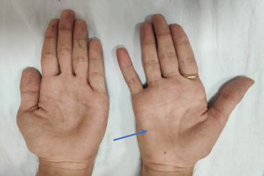
Degeneration of LMNs causes fasciculation, cramps, muscle atrophy and marked weakness, which is often more problematic for patients than the spasticity, hyper-reflexia and modest weakness associated with loss of UMNs. Babinski and Hoffmann signs, along with emotional lability are also typical of UMN degeneration.
ALS begins in the limbs in about two-thirds of patients, most often in the arms. The first symptoms are usually unilateral and focal. Early findings include foot drop, difficulty walking, loss of hand dexterity or difficulty lifting the arms over the head. Eventually, limb function can be lost, leading to dependence on caregivers. Patients may fall or lose the ability to walk all-together. Bulbar-onset disease, often occurring in older women, appears to have worse prognosis.22 Dysarthria usually begins before dysphagia; symptoms may progress to anarthria, drooling and malnutrition. An atrophied fasciculating tongue is so characteristic of bulbar ALS that it is virtually diagnostic of the condition. Axial weakness can cause dropped head and kyphosis, features associated with pain and poor balance. Sphincter and sensory functions are usually spared. Eye movements are preserved until advanced stages.
Cognitive impairment in ALS was first described by Pierre Marie in the 19th Century,23 but the association was considered uncommon until recently. Overt frontotemporal dementia occurs in approximately 15 %of patients, but as many as 50 % are impaired by neuropsychological tests.24,25 Changes involve language, judgment, personality, affect and executive function. Patients with ALS and dementia have shorter survival, possibly as a result of poor decision-making ability.26 Depression and anxiety can occur during any stage, from diagnosis to the time of respiratory failure, though patients suffering from ALS often approach the disease philosophically and rates of depression may be lower than expected.27 When present, emotional symptoms impair quality of life through poor appetite and sleep, and feelings of hopelessness. Pain can sometimes result from degeneration of sensory neurons, and more commonly from contractures, loss of mobility, inability to turn in bed, or bedsores. The suffering from being unable to move can be extreme.28 Morning headache, weakened cough, orthopnoea and exertional dyspnoea are early respiratory symptoms. Later, shortness of breath develops, first during simple tasks such as dressing and eating, and eventually at rest. The diagnosis depends on progressive UNM and LMN findings by history and examination. Electromyography confirms widespread LMN disease and excludes other conditions such as multifocal motor neuropathy with conduction block. Brain and spinal MRI exclude conditions that affect the UMN such as cervical spondylosis. Occasionally the brain MRI shows bilateral signal changes in the corticospinal tracts, a finding that is pathognomonic of ALS.
The El Escorial criteria help standardise the diagnosis for clinicalresearch studies (see Table 1).29 Progressive LMN disease by clinical and electromyographic examination, and clinical UMN signs are the core. Patients are classified by the number of involved body regions: bulbar,cervical, thoracic or lumbosacral. Recent modifications created on Awaji Island near Japan may improve diagnostic sensitivity, particularly for those with bulbar-onset in whom limb findings can be subtle.30
Pathogenesis
More than a century after the original descriptions by Charcot, the aetiopathogenic mechanisms are undiscovered for most patients. Approximately 5–10 % of ALS is familial. Mutations have been identified in some forms, but the mechanisms by which gene alterations cause ALS await elucidation. The first mutation discovered was in the superoxide dismutase Cu/Zn cytosolic (SOD1) gene on chromosome 21.31 The SOD1 mutation was used to create an animal model of ALS that has provided better understanding of disease physiology and served as an important means to screen new drugs. Currently, 15–20 % of fALS cases, mostly autosomal dominant, are due to one of more than 150 mutations in the SOD1 gene. Five percent of sporadic forms carry a similar mutation. Three other genes are linked to fALS: TAR DNA-binding protein 43 (TARDBP), fused in sarcoma (FUS/TLS) and C9ORF72, which may be the most common.32
There are a few leads for the causes of sALS. An elevated estimate of heritability in twin studies;33 gender predominance depending on phenotype;34 and familial aggregation of neurodegenerative disorders35,36suggest a genetic contribution. Genome-wide association studies (GWAS) have provided clues but no clear genetic explanations, however.37–41
GWAS are difficult to conduct in ALS because sample sizes must be very large to detect small genetic effects.42
Advancing age and exposure to tobacco smoke are associated with ALS,43 but there is currently little evidence to support the contribution of other environmental risk factors.43–48 A pooled analysis of five large cohorts found modest but significant associations with active smoking, former smoking, duration of smoking and quantity of cigarettes.49 Other possible associations include athleticism,50 particularly professional sports, pesticide exposure51 and service in the first Gulf war.52 A study in Italy from 1970–2001 found a 6.5-times higher risk in soccer players than non-players.53 Trauma is a possible contributor to all neurodegenerative diseases.54 A viral hypothesis, based on the analogy to enteroviral poliomyelitis, has existed unproved for decades; neuropathological studies and viral cultures have not shown evidence of viral attack.55 Associations with electrical fields and various toxins are also uncorroborated.56
Despite the absence of known causes, more is known about the physiology after disease-onset. Mechanisms that contribute to cell death include mitochondrial dysfunction, protein aggregation, generation of free radicals, excitotoxicity, disrupted axonal transport, inflammation and apoptosis.,sup>57 Nogo-A, a protein that inhibits regrowth of axons, is increased in the muscle of patients and in the murine model of ALS.58,59 Cell-to-cell transmission of aggregated misfolded protein, common to all neurodegenerative disorders, may be responsible for the spread of the disease in the brain.60
Management
There is no cure yet for ALS, so care is aimed at maintaining quality of life and prolonging life as much as possible. The current foundations are a single neuroprotective medication, multidisciplinary clinics and ventilatory support. Some therapies can help relieve symptoms.
Riluzole
Riluzole was developed because it possesses anti-glutamatergic properties; it might act in ALS to reduce exitotoxicity and apoptosis. Riluzole slowed progression of ALS in two randomised controlled trials.20,61 In the first trial, riluzole reduced mortality by 38.6 % at 12 months and 19.4 % at 21 months. In the second trial, the adjusted risk of death of 100 mg riluzole per day was 0.65 (p=0.002) compared to placebo. A meta-analysis of three trials that included 876 riluzole-treated and 406 placebo-treated patients showed that riluzole prolongs survival by approximately 11 %, or about two months.62 The most common side effects are fatigue, somnolence, nausea, diarrhoea and dizziness. Elevation of liver enzymes can occur, but rarely to levels that are clinically meaningful. Most patients in Europe, where the health systems cover the cost and more than half of patients in the US, take riluzole.63
Multidisciplinary Care
Multidisciplinary clinics were developed for ALS in the 1980s. A standardised approach was instituted in France in 2002 and most large centres in developed countries currently offer multidisciplinary care.64 Consensus publications guide diagnosis and treatment.65,66 Patients treated at multidisciplinary clinics may have higher quality of life67 and longer survival.17
Multidisciplinary care is orchestrated by a neurologist. Thorough evaluations, usually done every 3–6 months, by a team ensure that impending problems are handled efficiently. Patients and families see professionals from different disciplines in one sitting, conserving energy and time. Multidisciplinary teams also see many patients with ALS, so their level of experience is high. Care is focused on education and support, helping patients to make decisions about treatments. Discussions include advanced directives, treatments for nutritional and respiratory insufficiency and research.64 Advanced directives allow patients to make educated decisions about life support and help guide the multidisciplinary team in setting goals for care. Team members and a standard approach to care are outline in Table 2.
Additional staff available to clinics may include neuropsychologists, respiratory therapists, a pulmonologist, a gastroenterologist, a psychiatrist and an orthotist. Using the evidence gathered by the various professionals, the neurologist synthesises a plan for the patient. The summary and recommendations are conveyed to the patient’s primary physician so that care continues in tandem.
Nutrition and Gastrostomy
Inadequate nutrition and dehydration become common as ALS advances. The prevalence of undernourishment ranges between eight and 54 %,58,69 and is multifactorial. Bulbar muscle weakness and dysphagia, inability to eat adequately due to arm weakness and hypermetabolism all contribute to negative calorie balance. Reduced lean body mass and less physical activity accentuate muscle turnover. The end result is weight loss, which can be rapid and lead to more rapid clinical deterioration.70
Monitoring weight is the simplest way to assess caloric balance in the clinic. Calculation of body mass index (BMI) using height and weight is also used. Recommendations vary according to stage: initially, adaptations in food texture and postural changes such as the chin tuck are adequate. Nutritional supplements can be tried once oral intake becomes difficult. When positive caloric balance is no longer possible orally, a gastrostomy is indicated.
While gastrostomy ensures adequate nutrition, beneficial effects on quality of life and life expectancy have not yet been demonstrated.70,71 Some patients elect to have gastrostomy too late in the disease course to receive meaningful benefit.72 The ideal timing for gastrostomy awaits definition, but practice guidelines suggest that the procedure is safer when the vital capacity is above 50 % of predicted.69,73 Parenteral supplementation can be tried for those too ill to receive gastrostomy.74
Ventilatory Support
As respiratory muscle weakness advances, patients develop symptoms of dyspnoea, orthopnoea, sleep fragmentation, daytime fatigue and morning headaches. A weakened cough due to diaphragmatic and bulbar muscle weakness can lead to excess secretions, poor airway clearance and aspiration. The history, physical examination, overnight pulse oximetry and vital capacity (VC) are standard assessments that are done serially. The maximal inspiratory and expiratory pressures (MIP and MEP) correlate with respiratory muscle weakness.73 A MIP of <60 cm H2O is a predictor of reduced survival. Sniff nasal inspiratory pressure (SNIP), a noninvasive measure of inspiratory force, estimates intrathoracic pressure and is sensitive to respiratory muscle weakness. SNIP declines predictably over time in ALS patients and predicts survival time. A transcutaneous carbon dioxide sensor can detect carbon dioxide levels due to muscle weakness.75 The cause of death in ALS is usually respiratory, with approximately 60 % of patients having a predictable gradual decline in function and 35 % having sudden death.76
Noninvasive ventilation (NIV) is the standard intervention for patients with respiratory insufficiency.64 The bi-level intermittent positive-pressure ventilator, which is triggered by the patient’s inspiratory efforts and shuts off during exhalation, facilitates physiological function. When used at least 4 hours per day, NIV reduces the work of breathing, improves gas exchange, enhances sleep quality,77 extends survival,78 and may improve cognition,79 as well as lessen weight loss. Guidelines for use and timing of OF NIV73,80 are summarised in Table 3.
Oxygen is usually prescribed with NIV to prevent inhibiting respiratory drive in the setting of elevated serum carbon dioxide levels.
Invasive ventilation with tracheostomy is chosen by fewer than 5 % of patients.76,81 Invasive ventilation probably extends survival but requires 24-hour supervision and is costly. In long-term survivors, the disease might progress to the point where they can no longer communicate even using eye movement. Some patients choose to have respiratory support withdrawn, a decision that is ethical as long as opiates and anxiolytics are prescribed to avoid suffering when the ventilator is disconnected.
An assisted cough device, suction machine, theophylline, antibiotics, mucolytics, and expectorants can reduce symptoms.82 Pneumonia and influenza vaccines reduce pulmonary infections.73
Symptomatic Agents
Diverse medications, most used off-label, can reduce symptoms due to ALS (see Table 4). Some treatments improve quality of life and a few appear to extend life.
Palliative Care
All of ALS care is palliative because of the intransigently progressive course. Since publication of international recommendations for NIV, systematic evaluations have made discussions more matter-of-fact, but end-of-life decision making is complex and communication is key to a dignified and comfortable transition.
As in many chronic diseases with fatal outcomes, palliative care such as hospice can provide a framework for decisions about life support.
The goal of much of ALS care is to avoid suffering. Medications to relieve suffering, including anxiolytics and opioids, can be prescribed under the direction of a hospice team. Narcotic medications are effective for treating pain, nocturnal discomfort and breathlessness long before the terminal phase of the illness.83 End-of-life palliation is usually done at home, but inpatient hospice wards exist for patients who do not wish to die at home. Hospice teams provide symptom management through the use of medications and emotional support for patients and families.
Current Clinical Trials
Since two randomised controlled trials showed the effectiveness of riluzole and its approval in 1996, numerous drugs have been tested with negative results.84 This unbroken line of unsuccessful trials over 18 years despite advances in other areas of ALS research has raised important questions. Problems in preclinical models and trial design account for some of the difficulties.15 Recommended methodologies for animal studies85 and explanations for negative human trials86 are now published. ALS is a complicated disease to study; the disorder is rare, heterogeneous and disabling, correct doses are difficult to identify, outcomes are clinical and have high variance, and progressive weakness can lead to missing data.15 Investigators are searching for biomarkers so that diagnosis and progression measurement are more efficient.87
New trials are targeting muscle proteins, genes and gene products; utilising cell replacement therapies; and seeking ways to stabilise energy expenditure. Antagonsim of Nogo, a muscle protein that inhibits neurite outgrowth, may enhance reinnervation and is being tested in ALS.88 Stem cell therapies are also being tried.89 Cell therapy strategies utilise various types of stem cells to study disease pathophysiology, support neurons and surrounding cells through release of neurotrophic factors, or directly replace cells. Possible sources of stem cells for ALS include bone marrow, mesenchymal stem cells, neural stem cells, astrocyte precursor cells, and induced pluripotent stem cells. Energy depletion also appears to influence the development of the disease and is associated with worse prognosis.69 Two clinical trials evaluating nutritional interventions from complementary approaches are underway. The first trial is assessing a high fat diet given by tube feedings. The second trial is examining the safety and efficacy olanzapine, a drug that promotes weight gain. Gene therapy approaches, including antisense-mediated vectors, are also being tried to silence causative genes like SOD-1 or other molecular targets.’
Future Directions
As in other neurodegenerative disorders, affected neurons in ALS contain aggregated protein inclusions. The focal onset and contiguous spread of the disease clinically is mirrored by the pathology. Abnormal protein is localised at first and then spreads to reach other areas of the brain. In ALS, the inclusions occur first in motor neurons. In patients with ALS and dementia, the inclusions are found throughout the frontal and temporal lobes. Some of these proteins may have prion-like domains, with a propensity to self-aggregation. Abnormal proteins might induce normal protein to change conformation, causing the disease to move from diseased to healthy cells, migrating through the brain. Protein aggregates are themselves associated with other pathophysiological processes such as mitochondrial dysfunction and energy depletion, glutamate excitoxicity, and induction of inflammatory mediators. Misfolded protein might be a target for therapeutic intervention, which could be applicable to other neurodegenerative diseases.60 A therapeutic breakthrough in one neurodegenerative disorder would likely translate rapidly to other disorders.
Conclusions
ALS is still a mysterious disease with few known aetiologies or meaningful treatments. The course is the most rapid of any neurodegenerative disorder. Care is multidisciplinary and includes respiratory support, supplemental feeding and riluzole, which appear to extend survival modestly. Symptomatic medications can improve quality of life, but more need to be tested in trials for ALS. Numerous trials have targeted pathophysiological processes, but there have been no recent successes. The important overlap between ALS, frontotemporal dementia and other neurodegenerative conditions marked by accumulations of misfolded protein is being studied with increasing earnest. The spread of the disease clinically and pathologically is reminiscent of local seeding by prion proteins. Therapeutic strategies might include drugs to slow the formation of misfolded protein or aid in its dissolution. Meanwhile, patients are offered modern care that can ensure comfort and dignity during this most difficult disease.


