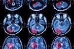
The classical perception of Parkinson’s disease (PD) heavily emphasises its motor aspects, cognitive features and dementia associated with the disease being largely ignored. Epidemiological studies performed in the last few decades revealed the substantially high incidence and prevalence of cognitive impairment and dementia in PD. Clinical studies using detailed neuropsychological and behavioural assessments have helped to understand its cognitive and behavioural profile, while clinical– pathological correlation studies have revealed the underlying pathology and ascertained the delineation of dementia associated with PD (PD-D) as a distinct entity. This article provides an update on the epidemiological, clinical and pathological features of PD-D and recent treatment efforts.
Epidemiology
Dementia associated with PD has been increasingly better recognised, probably because patients with PD survive for longer than before thanks to modern treatment. Although subtle cognitive deficits can be found in newly diagnosed patients with PD,1 dementia itself is strongly associated with advanced age and severe disease.2 In population-based, cross-sectional studies the prevalence of dementia has been reported to be 28–41%. A meta-analysis of 12 carefully selected studies revealed a cross-sectional prevalence of close to 30%.3 The incidence increases up to six-fold and the cumulative incidence over eight years of follow-up was described to be 78%.4,5 The longitudinal Sydney study revealed that 15 years after the diagnosis 85% of the patients had cognitive impairment, with 50% fulfilling criteria for dementia.6 The main risk factors include old age, severity of motor symptoms, akinetic–rigid form of the disease and subtle deficits in verbal fluency with executive functions and memory performance at baseline.
Clinical Features
The proto-typical dementia syndrome associated with PD (PD-D) has characteristic clinical features, which can be best summarised as a ‘dysexecutive’ syndrome, with prominent impairment of attention, visual–spatial dysfunction, moderately impaired memory and accompanying behavioural symptoms such as apathy and psychosis.8,9
Cognitive Features
Impaired attention is an early and prominent feature of patients with PD-D. Impairments in reaction time and vigilance, as well as fluctuating attention, are common findings, similar to the related disorder, dementia with ‘Lewy bodies’ (DLB). Compared with patients with AD, PD-D patients are more apathetic and have more prominent cognitive slowing, the magnitude of which is usually disproportionate to the general level of cognitive performance.
Impairment in executive functions is the core feature of PD-D. Deficits include poor performance in tasks involving rule-finding, problem-solving, planning, set elaboration, set shifting and set maintenance. Patients have more difficulty when they have to develop their own strategies; performance improves when external cues are provided. Abnormalities in executive functions occur early in the course of PD-D and are prominent throughout the course of the disease.
All memory functions are impaired, including working memory, explicit memory and implicit memory such as procedural learning. Deficits in working memory can be found early in the disease course. In the majority of patients, the relative severity and profile of memory impairment, compared with impairment in attention and executive functions, differ from those seen in AD. Compared with patients with AD, memory impairment is less severe and is characterised by a deficit in free recall with relatively preserved recognition, indicating that information is stored but not adequately accessed. Memory performance usually improves when semantic cues or alternatives are provided. Based on this profile, it was suggested that the impairment of memory in PD-D might be due to executive dysfunction, which causes difficulties in accessing memory traces due to a deficiency in developing appropriate search strategies.
Another characteristic feature of PD-D is early and prominent deficits in visual–spatial functions, in both perception and construction. These deficits are usually disproportionate to the overall severity of dementia. Compared with AD patients with a similar severity of dementia, PD-D patients perform worse in all perceptual scores, and those with visual hallucinations tend to have worse visual perception than those without.10 Visual–spatial abstraction and reasoning were more impaired in patients with PD-D, whereas visual–spatial memory tasks were worse in patients with AD. This was especially evident in more complex tasks that required planning and sequencing.11
In contrast, the other lateralised function, language, seems to be largely preserved in patients with PD-D. Core language functions are usually normal. Deficits consist of impaired verbal fluency and mild word-finding difficulties. Impaired verbal fluency, the main language impairment found in PD-D, is more severe than that seen in patients with AD. Other deficits include a decreased content of spontaneous speech and impairment in the comprehension of complex sentences, but these are to a significantly lesser extent than in patients with AD.12 As for impairments in other domains, it was suggested that most of the language deficits, such as impaired verbal fluency and word-finding difficulties, may not reflect a true involvement of language functions, but may be related to the executive dysfunction.9
Behavioural Features
Patients with PD-D have prominent behavioural and neuro-psychiatric symptoms. Almost all patients demonstrate changes in personality, such as retardedness, social withdrawal and apathy. The most common neuro-psychiatric symptoms are depression, hallucinations, apathy and anxiety.13 Hallucinations and delusions can follow treatment with dopaminergic agents; they occur more frequently in patients with dementia and may be a harbinger of incipient dementia. In one study 83% of those with PD-D were found to have at least one psychiatric symptom, as opposed to 95% of those with AD. Hallucinations were more severe in PD-D, whereas increased psychomotor activity – including aberrant motor behaviour, agitation, disinhibition and irritability – were seen more commonly in AD. In PD-D, apathy was more common in mild stages while delusions increased with more severe motor and cognitive dysfunction.14 Rapid eye movement (REM) sleep behaviour disorder is also a frequent feature in PD-D, similar to other synucleinopathies such as DLB and multiple systems atrophy (MSA), but unlike other degenerative diseases such as AD or progressive supranuclear palsy (PSP).15
Clinical–Pathological Correlations
Three types of pathological substrates have been suggested to underlie dementia in PD. These include: cellular loss in discrete subcortical nuclei, notably dopaminergic cell loss in medial substantia nigra and nuclei of the other ascending pathways; co-incident Alzheimer-type pathology; and Lewy body (LB) type pathology in limbic and cortical areas. There have been a number of clinical–pathological correlation studies supporting one or the other view.7 Since the beginning of 2000, several carefully performed studies have become available that used alphasynuclein antibodies to identify LB pathology (which is more sensitive than the traditional ubiquitin staining), and assessed LB- and AD-type pathologies in parallel. The results of these studies suggest that dementia in PD best correlates with LB pathology in limbic and cortical areas.16–19 AD-type pathology, such as plaques and, less so, tangles, frequently co-exist; they are less predictive of dementia and they rarely reach a severity to justify a pathological diagnosis of AD. PD-D can be designated as an LB-related dementia, as it is closely related to dementia with LB. Recent genetic findings that triplication of alpha-synuclein gene, the protein that is the main constituent of LB, resulted in familial PD with dementia further support the defining role of LB-type pathology.20 The hypothesis that pathological changes in PD follow an ascending order, sequentially involving cerebral structures as the disease progresses, with limbic and cortical involvement beginning later in the disease process, may explain why dementia usually develops relatively late in PD.21
Biochemical Deficits and Treatment of Parkinson’s Disease Dementia
Although deficits in almost all ascending mono-aminergic pathways have been suggested to be associated with PD-D, the most consistent findings in biochemical, anatomical and functional studies favour a cholinergic deficit. Morphologically, there is a prominent loss of cholinergic neurones in the nucleus basalis of Meynert (nbM),22 to a greater extent than in those with AD.23 This cellular loss is reflected by cholinergic deficits, as demonstrated by reduced cholinacetyltransfarase and acetylcholinesterase activity in nbM and in the cerebral cortex. The severity of these deficits correlates well with the severity of dementia.24,25 In a comparative study of patients with AD, DLB and PD, mean midfrontal choline acetyltransferase (chAT) activity was found to be markedly reduced in PD and DLB compared with normal controls and also with those with AD.26 Functional imaging studies using positron emission tomography (PET) scans and cholinergic markers revealed similar findings: compared with controls, mean cortical AChE activity was lowest in patients with PDD, followed by patients with PD without dementia and AD patients with equal severity of dementia.27 In addition to impairment in the basal fore-brain cholinergic system, PD-D is also associated with neuronal loss in the pedinculopontine cholinergic nuclei that project to structures such as the thalamus.28
These substantial cholinergic deficits led to the investigation of cholinergic enhancement strategy in patients with PDD using cholinesterase inhibitors (ChE-I). All commercially available ChE-I, including tacrine, donepezil, rivastigmine and galantamine, have been tried in this patient population in some form, mostly in open-label trials or as case series. These reports are summarised in a review by Aarsland and colleagues.29 Despite their methodological limitations with regard to study designs and small sample sizes, an overall evaluation of these studies suggests that treatment with ChE-I provides an improvement in cognition as well as behavioural symptoms in most patients, without having a detrimental effect on motor functions in the majority of patients.
The only large randomised controlled study in patients with PDD was performed with rivastigmine.30 This study, abbreviated as ‘EXPRESS’, recruited 541 patients with mild to moderate PD-D who were randomised to receive rivastigmine or a placebo in a ratio of two to one. Primary efficacy parameters included the Alzheimer Disease Assessment Scale-cognitive section (ADAS-cog) for the assessment of cognitive functions and the Alzheimer Disease Collaborative Study – Clinical Global Impression of Change (ADCS-CGIC) for the assessment of overall change in the clinical status from baseline. Secondary clinical efficacy parameters included mini mental state examination (MMSE) for screening and staging, ADCS-Activities of Daily Living scale (ADCSADL), Neuro-psychiatric Inventory (NPI), a computerised test battery for the assessment of attention (CDR power of attention tests) and two tests for the assessment of executive functions, verbal fluency and 10-point clock-drawing test. In addition to standard safety parameters, part three (motor section) of the Unified Parkinson Disease Rating Scale (UPDRS) was assessed for an objective evaluation of motor functions. Of the 541 patients entered in the study, 410 completed and 131 patients prematurely discontinued. There were more discontinuations in the rivastigmine group (27.3 versus 17.9% under placebo); this was also the case for discontinuations due to adverse events (17.1 versus 7.8% under placebo). Both primary efficacy end-points showed statistically significant improvements in favour of rivastigmine. ADAScog patients on rivastigmine showed a 2.1 improvement at 26 weeks, whereas patients on placebo deteriorated by 0.7 points (p<0.001). The mean scores for the seven-point ADCS-CGIC at week 24 were 3.8 in the rivastigmine and 4.3 in the placebo group (with a score of 4 indicating no change, lower scores indicating improvement and higher scores indicating worsening from baseline). Comparison of outcomes across all response categories revealed a statistically significant difference in favour of rivastigmine (p=0.007). More patients on rivastigmine showed an improvement (40.8 versus 29.7% on placebo) and more patients on placebo deteriorated (42.5 on placebo versus 33.7% on rivastigmine). On all secondary efficacy parameters there were statistically significant differences in favour of rivastigmine. Neuro-psychiatric symptoms as measured with NPI showed an improvement on rivastigmine and no change from baseline on placebo.
There were improvements in the 10-point clock-drawing test, verbal fluency and power of attention and MMSE scores on rivastigmine, whereas patients on placebo worsened compared with their baseline scores. On ADCS-ADL patients on rivastigmine showed a minimal worsening, whereas those on placebo did so more significantly. Adverse events were significantly more frequent on rivastigmine. The main adverse events were those related to the gastrointestinal system. Nausea and vomiting were the most frequent (29.0 versus 11.2% nausea, and 16.6 versus 1.7% vomiting on rivastigmine and placebo, respectively). Worsening of parkinsonian symptoms was more frequently reported as an adverse event on rivastigmine (27.3 versus 15.6% on placebo), mainly driven by a worsening of tremors (10.2% on rivastigmine versus 3.9% on placebo). The objective measures of motor symptoms as assessed by UPDRS part three did not reveal any significant differences or trends between the two treatments. There were no clinically relevant changes on vital signs, bodyweight and ECG or laboratory parameters. On the basis of the ‘EXPRESS’ study, rivastigmine became the first ChE-I to be granted a marketing approval for patients with mild to moderate PD-D.
Conclusions
Dementia affects 30–40% of patients with PD cross-sectionally; some form of cognitive impairment develops in 85% of patients 15 years into the diagnosis of their disease. Characteristics of dementia associated with PD include prominent impairments in attention, executive and visual–spatial functions and, to a lesser degree, in memory. Behavioural symptoms such as depression, hallucinations, delusions, apathy and anxiety are common. The main pathology associated with PD-D is LB in limbic and cortical areas. Biochemically, the most prominent deficits are cholinergic, and treatment with ChE-I may provide benefits in cognitive and behavioural symptoms without an undue worsening of motor symptoms. The only existing randomised controlled study is for rivastigmine. ■


