Clinical Features of Progressive Supranuclear Palsy
Clinical Features of Progressive Supranuclear Palsy
Progressive supranuclear palsy (PSP) is a neurodegenerative disorder characterised by parkinsonism, supranuclear ophthalmoplegia, dysphagia and cognitive dysfunction. The National Institute of Neurological Disorders and Stroke (NINDS) PSP criteria are widely used for clinical diagnosis.1 However, the clinical phenotypes of pathologically confirmed PSP patients are heterogeneous. Recently, PSP has been clinically classified into two phenotypes: Richardson’s syndrome and PSP-parkinsonism.2 Those patients who have Richardson’s syndrome are neurologically characterised by the classic features, including the early onset of postural instability and falls, supranuclear vertical gaze palsy and cognitive dysfunction. PSP-parkinsonism is frequently misdiagnosed as Parkinson’s disease in its early clinical stage and is characterised by asymmetrical onset, tremor and moderate initial therapeutic response to levodopa. Differentiating between Richardson’s syndrome and Parkinson’s disease is not difficult, but differentiating between PSP-parkinsonism and Parkinson’s disease is occasionally challenging. Early differentiation of these diseases is necessary because the therapeutic strategy and outcome are substantially different (e.g. PSP patients have much worse prognosis).
Diffusion-weighted Imaging
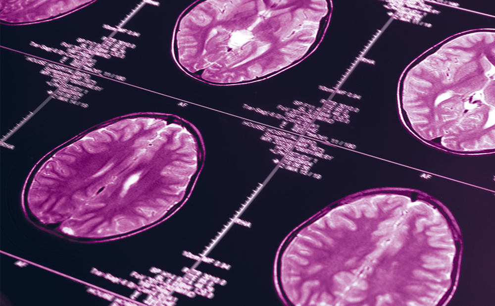
Magnetic resonance imaging (MRI) is a promising non-invasive tool that supports or complements clinical diagnosis. It is also useful for the investigation of the pathophysiology of diseases. Recently, several new MRI techniques have been developed, and one of the most important of these tools is diffusion-weighted imaging,3 which can be used to measure the diffusion of water molecules in the brain.4 Brain diffusion is influenced by tissue density, neuronal axon and vessel direction and capillary flow. The apparent diffusion coefficient (ADC) or mean diffusivity (MD) is a parameter that represents diffusion intensity, and high ADC or MD values indicate increased diffusion intensity (see Figure 1). Diffusion intensities along three orthogonal directions are represented as λ1, λ2 and λ3, and MD values are calculated as [λ1, λ2, and λ3]/3. Axial diffusivity (λ1) and radial diffusivity ([λ2 + λ3]/2) are occasionally used as additional measures to identify whether differences in anisotropy may be caused by diffusion parallel or perpendicular to the white-matter fibres, respectively. Cytotoxic oedema is a typical pathological condition in which decreased ADC values are indicative of cerebral infarction5,6 or other conditions. ADC values are increased by vasogenic oedema7 or demyelination.8 In neurodegenerative diseases, ADC values are increased in affected areas because of disruption of neuronal axons.9,10
Diffusion Tensor Imaging
Diffusion tensor imaging was introduced as a new imaging modality in 1994.11 Molecules inside neuronal axons have a low probability of crossing the myelin membrane, and thus brain diffusion in this modality is usually anisotropic. Fractional anisotropy (FA) provides an index of brain anisotropy; the FA values are calculated as a value from zero to one, with a score of one representing tissue that is more anisotropic (see Figure 2).
The posterior limbs of the internal capsule, cerebral peduncles, corpus callosum, optic radiations and middle cerebellar peduncles are representative anisotropic tissues, whereas the grey matter, including the cerebral cortices and basal ganglia, are less anisotropic (see Figure 2).12 The development of diffusion tensor imaging enables brain diffusion to be measured in multiple directions (see Figure 3) and the FA in each direction to be calculated for each voxel.13 Diffusion tensor tractography is a recently developed method to visualise the neuronal fibres in 3D by computing the connections of diffusion tensor in adjacent voxels (see Figure 4).14
Measuring Diffusion Properties
Two approaches may be used to measure and analyse diffusion properties such as ADC and FA values. One approach is to measure diffusion properties by setting the regions or voxels of interest, which are classically defined as circular or square regions, in each subject. Regions of interest can be set along the tractography (‘tract of interest’), and the diffusion properties in entire voxels on a particular neuronal pathway can be measured.15 The other approach is to perform voxel-by-voxel analysis of the diffusion properties. The statistical parametric mapping method is an algorithm that is generally used in neuroimaging studies, and whole brain analysis can be performed without the subjective dispersion of setting regions of interest.16 Appropriate normalisation, segmentation into white matter and smoothing of diffusion images on specific software are necessary for performing voxel-by-voxel diffusion analysis.
Diffusion Tensor Abnormalities in Progressive Supranuclear Palsy
Supratentorial Structures
Padovani et al. were the first to perform diffusion tensor analysis in patients with early PSP.17 They conducted voxel-by-voxel analysis of diffusion tensor images using statistical parametric mapping, combined with grey-matter analysis using optimised voxel-based morphometry.18 Their study revealed decreased FA values in the superior longitudinal fasciculus, anterior part of the corpus callosum, arcuate fasciculus, posterior thalamic radiations and internal capsule (see Table 1), in addition to decreased FA values in the affected grey matter, including the premotor cortex, frontal operculum, anterior insula, hippocampus, and parahippocampal gyrus, the pulvinar, dorsomedial and anterior nuclei of the thalamus and the superior and inferior colliculus.
These results are suggestive of frontal dominant grey- and white-matter cerebral damage, and are compatible with previous diffusion and morphometric studies.19,20 Nilsson et al. reported a visual reduction of cortical projection fibres in patients with PSP in 3D diffusion tensor tractography on 3 Tesla MRI.21
Recently, we analysed the corpus callosum as a representative area of cerebral commissure fibres by diffusion tensor imaging22 using a new partitioning method for the human corpus callosum.23 The results showed that the FA values were significantly decreased in CC1 (the prefrontal area) and CC2 (the premotor and supplementary motor areas), and the ADC values were significantly increased in CC1 and CC2. Receiver operating characteristic analysis showed excellent reliability of FA and ADC analyses of CC1 for differentiating PSP from Parkinson’s disease. Although FA values generally decrease and ADC values increase during the normal ageing process in the frontal white matter and anterior corpus callosum,24–29 our results showed differences in diffusion parameters between age-matched PSP patients and normal subjects. Our results suggest that the frontal commissure fibres are preferentially damaged in patients with PSP, and thus diffusion tensor imaging of the corpus callosum is helpful for the diagnosis of PSP.
Infratentorial Structures
Blain et al. reported diffusion changes in the brainstem in patients with PSP and multiple system atrophy using a region-of-interest approach.30 They revealed increased MD values in the decussation of superior cerebellar peduncles in patients with PSP, in contrast to decreased FA values in the middle cerebellar peduncles and increased MD values in the pons in patients with multiple system atrophy (MSA). Nilsson et al. also showed the same distribution of diffusion abnormalities in patients with PSP and MSA using diffusion tensor tractography on 3 Tesla MRI.21 These results showed that cerebellar efferent fibres (superior cerebellar peduncles) are preferentially affected in patients with PSP, whereas cerebellar afferent fibres (pons and middle cerebellar peduncles) are damaged in patients with multiple system atrophy.
Clinical Application and Limitations of Diffusion Tensor Imaging in Progressive Supranuclear Palsy
Diffusion tensor imaging is a feasible method for investigating human white-matter conditions. However, it is not fully practical for differentiating PSP from other parkinsonisms such as Parkinson’s disease or multiple system atrophy because there are some overlaps of diffusion parameters between PSP and other forms of parkinsonism.22,30 Analysis using statistical parametric mapping is not applicable for individual cases unless the Z score of diffusion parameters can be calculated for each selected area, and this requires specialised software and training.17 In visual inspection of diffusion tensor tractography images,21 it is difficult to set a cut-off line of abnormalities of fibre visualisation. In summary, diffusion tensor analysis is not essential for the diagnosis of PSP, although it can be an additional test for further confirmation of the diagnosis.
Future Perspectives of Diffusion Tensor Analysis in Progressive Supranuclear Palsy
To the best of my knowledge, there has been no detailed correlation analysis between clinical features and diffusion tensor parameters in PSP. Frontal diffusion abnormalities probably reflect clinical frontal lobe dysfunction in conceptualisation, mental flexibility, motor programming, sensitivity to interference, inhibitory control and environmental autonomy for executive functions. Diffusion tensor analysis regarding supranuclear ophthalmoplegia, which is a core feature of PSP, is also necessary for further understanding of the pathophysiology of PSP.
Conventional diffusion tensor imaging using echo planar technique is generally insufficient for detailed analysis of the brainstem because of a strong susceptibility effect around the skull base. Diffusion tensor imaging using a line scan technique31 may be feasible for analysing the brainstem in patients with PSP.32,33 Finally, the clinical subtypes of PSP, Richardson’s syndrome and PSP-parkinsonism2 have not yet been investigated by diffusion tensor imaging.
Early differentiation of these subtypes of PSP is important for planning a clinical strategy because the treatment responses and disease outcome are different. Diffusion tensor imaging is a feasible method for investigating brain conditions in PSP. ■